Shared equipment
- Top
- Shared equipment
- Scanning X-ray Photoelectron Spectroscopy
Shared equipment
Scanning X-ray Photoelectron Spectroscopy
Scanning X-ray Photoelectron Spectroscopy
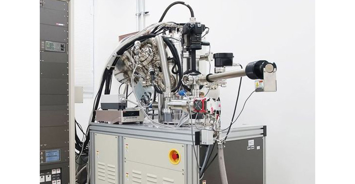
PHI5000 VersaProbe II
Equipment Overview (Purpose)
Through the irradiation of a solid sample with X-rays, the spectrum of photoelectrons emitted from a region of about 10 nm in depth on the surface can be measured, and the composition and chemical bonding state of the surface can be analyzed.
Main Features and Applications
【Features】
- Analysis by scanning microfocus X-ray source
The X-ray beam diameter can be selected from 10μm to 200μm, enabling the qualitative and the semi-quantitative analysis of the sample surface, as well as the analysis of the chemical bonding state. In addition, composition mapping and chemical bonding state mapping can be performed for areas of up to 1 mm x 1 mm. -
SXI (Scanning X-ray Image) for precise analysis location
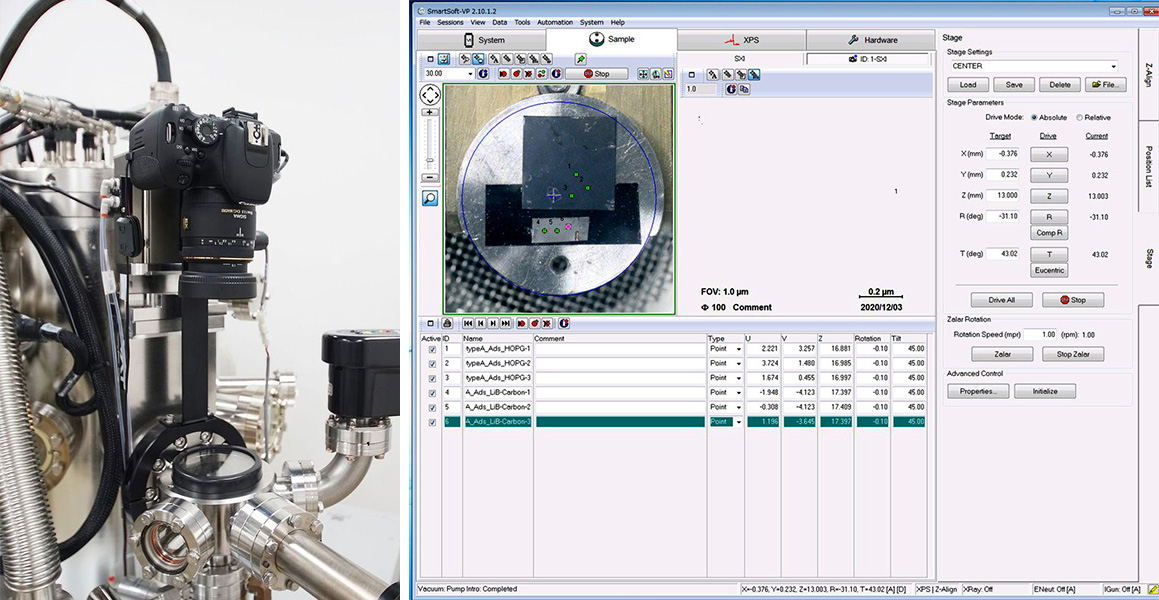
Microfocused X-rays are scanned over an area of up to 1.4 mm x 1.4 mm, and the excited photoelectrons are captured via an electron analyzer to obtain a two-dimensional image (SXI image) that reflects the sample shape and composition information. Because the photoelectron image excited using the same X-rays as in the actual analysis is used, it is easy to identify and quickly confirm the precise analysis position. - Analysis of insulating materials using a dual neutralization gun
The simultaneous irradiation of low-energy electrons and Ar ions enables the analysis of the chemical bonding states of insulating materials. - Automatic multi-point analysis
Automatic multi-point analysis can be performed over a long period according to preset measurement points and conditions.
【Applications】
- Bonding state analyses of metals, oxides, and nitrides
- Surface functional group and bonding state analyses of carbon materials (graphene, DLC, CNT) and catalyst materials
- Composition and bonding state analyses of organic materials and polymers
- Bonding state analyses of cathode and anode materials for lithium-ion batteries via non-exposure to the atmosphere
- Depth analysis via argon sputtering and angle-resolved measurement
Main Specifications
| Minimum beam diameter | φ10μm | ||||||||
|---|---|---|---|---|---|---|---|---|---|
| Detector | 16 channel (Interlace mode supports 128 channels) |
||||||||
| X-ray scan range | Maximum 1.4mm×1.4mm(continuously variable) | ||||||||
| Ion gun acceleration voltage | 0~5kV (SiO2 equivalent sputtering rate:0.4~11 nm/min) | ||||||||
| Sensitivity/resolution (Conditions: 20μm beam diameter, Ag) |
|
| Ion gun cluster range | 2mm×2mm、3mm×3mm |
|---|---|
| Ultimate vacuum | 6.7×10⁻⁸Pa or less |
| Maximum energy resolution | 0.5eV以下 (Ag3d 5/2) |
| Maximum sensitivity | 1,000,000cps (Ag3d 5/2 half-value width 1.0eV) |
| Monochromator Roland diameter | 200mm |
Available Optional Features & Option Purpose
| Available Optional Features | Option Purpose |
|---|---|
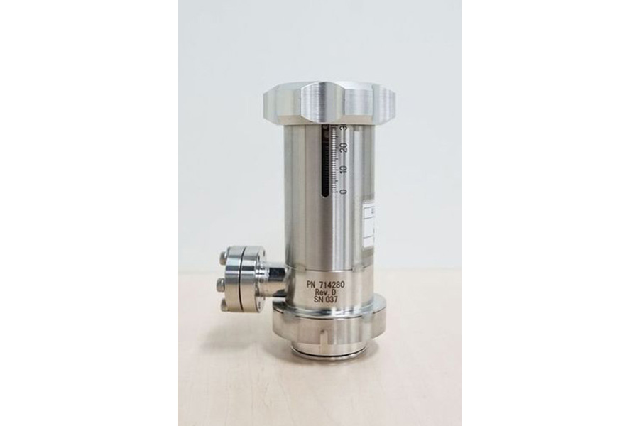
| Available Optional Features:Transfer Vessel | Option Purpose:For highly active materials that react with atmospheric components, this feature makes it possible to maintain the materials’ original states by introducing them into the system without atmospheric exposure. |
Equipment Photo
Click on the photo to enlarge.
- Tohoku University
- Institute for Materials Research, Tohoku University
- Institute of Fluid Science, Tohoku University
- Institute of Multidisciplinary Research for Advanced Materials, Tohoku University
- Research Institute of Electrical Communication, Tohoku University
- Head Office of Enterprise Partnerships, Tohoku University
- Core Facility Center
- Bulk Soft Magnetic Materials, Tohoku University
- Advanced Imaging and Modeling Center for Soft-materials
(Tohoku AIMcS)
2013 © Material Solutions Center, Tohoku University. All rights.


